Spinal cord (back) pain can occur in any age, size, or breed of dog or cat.
- The causes of such pain can be caused by a number of issues including, but not limited to, intervertebral disc disease, trauma, infectious disease, or congenital problems.
- These conditions can include the musculature, vertebral bodies, tendons and ligaments, spinal cord, or a combination of several.
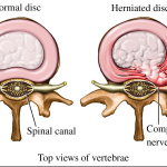
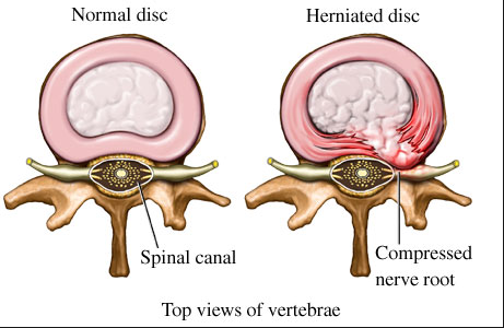
- The most common encountered issue resulting in back pain seen clinically is intervertebral disc disease (IVDS).
Clinical Signs
The most common breed affected by IVDS are the smaller breeds:
- Dachshund
Click to Enlarge
- Lhasa Apso
- Poodle
- Cocker Spaniel
- Shitzu
- Pomeranian
Signs
- Crying out in pain while try to jump, run, or play.
- Reluctance to jump or climb stairs normally.
- Hindlimbs weak or unsteady when trying to walk or stand.
- Paralysis of one or both hind limbs.
- Loss of bladder or bowel control
Diagnosis of IVDS
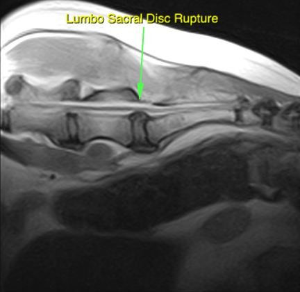
- An MRI is the best way to determine the location and the severity of the injury in your pets spine.
- This technology allows us to achieve greater visualization of spinal cord disease over the older techniques such as myelograms.
- Since we have an onsite MRI machine, this gives us better diagnostic capability and surgical precision when dealing with spinal cord injury.
- All spinal MRI’s are submitted and reviewed by a board certified radiologist prior to any surgical intervention.
-
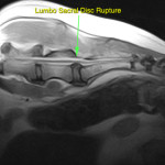
- Lumbo-Sacral Disc Rupture
-
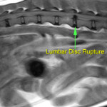
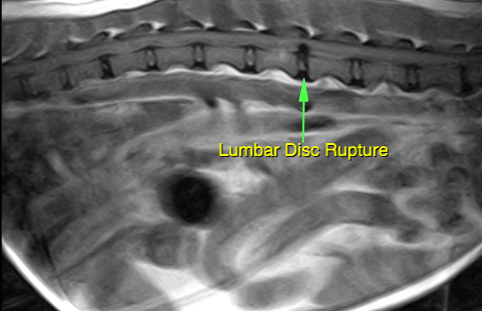
- Lumbar Disc Rupture
-
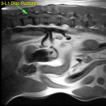
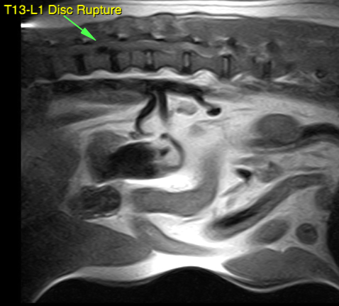
- T13-L1 Disc Rupture
Treatment
- Conservative therapy can result in resolution of clinical signs, however, if the pet does regain the ability to walk again, usually the function is not as good as it would be if surgery was performed.
- Surgery is frequently the best treatment.
- A surgical procedure called a hemilaminectomy involves removing a portion of bone from one side of the back bones at the level of the herniated disc .
- After the spine has been opened, the herniated disc material is removed from beneath the spinal cord.
- A procedure called fenestration may also be done to help prevent a future disc rupture. This involves opening the side of adjacent discs and removing the gelatinous nucleus pulposus material.
Potential complications
- Anesthetic death is a very uncommon complication.
- Infection of the spine, although possible, is very uncommon.
- Bladder infection may result from inadequate emptying of the bladder due to neurological dysfunction. Antibiotics are needed to correct this problem.
- Permanent paralysis can occur, but is uncommon.
- Side effects due to medications may include vomiting, diarrhea and loss of appetite. If any of these clinical signs occur notify our clinic or your routine care veterinary clinic.
- Leakage of air into the chest, which can be life-threatening, may occur if the thoracic vertebrae are being operated and the surgeon penetrates the chest cavity.
Postoperative care
- Medications that you may need to administer to your pet may include:
- Steroids: help reduce swelling and inflammation
- Muscle relaxants: help to minimize painful muscle spasms
- Analgesics: a variety of medications are available for this purpose
- Rest: Your pet should be kept in a crate at all times except for urination and bowel movements for a period of one month.
- Rehabilitation therapy is very important of patients that are weak or paralyzed.
- Passive rehabilitation: flex and extend the joints of the hindlimbs. These exercises should be done three to four times daily, 20 minutes per session.
- Active rehabilitation: assist your pet in a standing position. When your pet gets tired and lies down, allow a short period of rest (30 seconds) and then get him/her back into a standing position.
- Bladder care is needed in most dogs that have had surgery for a thoracolumbar spinal cord lesion. If your pet is paralyzed, bladder control also may be lost. You may need to express the bladder so that it does not become too distended. Once the pet regains voluntary motor function of the hind limbs, the bladder function usually has recovered.
- Check the incision for signs of infection: redness, swelling, pain or discharge. Do not allow your pet to scratch or rub the incision. If needed the neck may need to be bandaged until the incision has healed.
- A soft padded bed is essential during the recovery phase to prevent the development of bed sores/ulcers. If your pet cannot turn him/herself you will need to turn him/her from side to side every four hours.
- An evaluation of your pet will be made about 2 weeks after surgery at our hospital or the referring veterinary clinic.
Prognosis
- Following spinal surgery, it is common that the neurological status may be the same or somewhat worse than prior to surgery. This is caused by the surgical manipulation of the spinal cord. In most cases this is a temporary set back and improves in a few days. Dogs that are paralyzed prior to surgery usually have a 3 to 6 week convalescent period before they can walk again. At this point they frequently are still unsteady on their hind limbs. By 2 months after surgery most dogs are able to ambulate very well. Improvement in the strength of the hind limbs can progress until the 6th to 9th month after surgery.
- Dogs that have had one disc herniation event are at greater risk than ordinary dogs for a repeat event.